
The study from Frontiers in Medicine, March 2025 presents the first paleopathological evidence for lepromatous leprosy in Bronze Age Oman (2500–2000 BCE), specifically at the site of Dahwa. This extends the known geographic range of ancient leprosy beyond South Asia, where the earliest skeletal evidence had previously been found. The presence of leprosy at Dahwa, a site with strong material and cultural ties to the Indus Valley and Mesopotamia, supports the hypothesis that pathogens like Mycobacterium leprae spread along Bronze Age trade routes (just as COVID spread in recent times). The finding aligns with genetic and archaeological evidence suggesting the pathogen’s spread was facilitated by human mobility and exchange networks. One might even say that it reinforces the pervasiveness and importance of these trade routes, for, as the authors write, "leprosy requires an extensive period of close contact for transmission and the latent period can be decades," (p. 1).
The connection of the site to the ancient Indus civilization is not trivial: "A large number of Indus pottery sherds (around 72% of the total sherds) indicate the first interaction of the Umm an-Nar people with the Indus people after 2500 BCE," (p. 2). Copper slag fragments suggest that this interaction may have had to do with copper mining in Oman. It was the funerary structures of these local Omani inhabitants that yielded some 180,000 bone fragments used in the analysis, with yielded "the first evidence of the pathogen outside of South Asia," (p. 5).
The research demonstrates the utility of micro-CT scanning for non-invasively diagnosing leprosy in highly fragmentary and commingled skeletal assemblages. Micro-CT allowed the authors to characterize microstructural changes in three archaeological maxillae, identifying lesions consistent with lepromatous leprosy-such as atrophy of the anterior nasal spine, alveolar resorption, deterioration of the piriform aperture margin, and atrophy of the nasal septum. In short, the analysis suggests methods with which other ancient facial skeletons can be read for signs of this ancient disease as well as methods for better reading the presence of leprosy in modern CT scanning which could be important given how hard it is to diagnose this disease and long its incubation period.
Various questions for future research arise from the paper: how might the presence of leprosy in Bronze Age Oman reshape our understanding of disease transmission along ancient trade routes? What other pathogens might have spread similarly, and what evidence would we need to detect them? What do the mortuary practices at Dahwa suggest about community attitudes toward individuals affected by leprosy? Is there evidence for social exclusion, care, or specific treatment of the diseased in burial contexts? Considering the evidence for leprosy’s ancient spread, how might this inform our understanding of the co-evolution of humans and M. leprae? Are there implications for why some populations developed greater innate immunity? As with all ground-breaking research, the raising for multiple new issues is to be expected and likely this work will continue to yield new insights into ancient trade networks and all that they involved besides material goods.
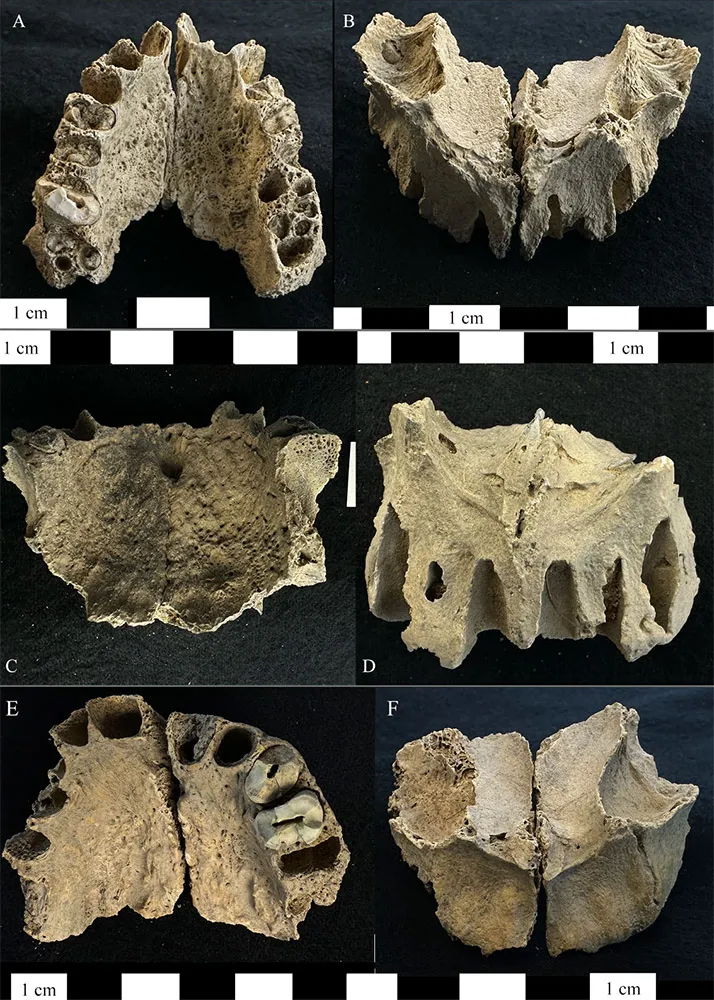
Images: Right and left maxillae from the bone pit at Dahwa included in this study. (A) Maxilla ID 012 inferior-dorsal view of the maxilla demonstrating periosteal new bone formation and abnormal porosity on the palatal surface. (B) The superior-anterior view of ID 012 showing porosity on the cortical surface of the maxillary sinuses, exposure of maxillary nerve V2, resorption of the anterior nasal spine and the margins of the pyriform aperture, and alveolar resorption of the anterior dentition. (C) Maxilla ID 093 inferior-dorsal view of the maxilla demonstrating periosteal new bone formation and abnormal porosity on the palatal surface as well as abnormal porosity on the mesial alveolar wall for the left first molar. (D) The superior-anterior view of ID 093 showing resorption of the anterior nasal spine, margins of the pyriform aperture, alveolar resorption around the anterior dentition. (E) Maxilla ID 044 inferior-dorsal view of the maxilla demonstrating periosteal new bone formation and abnormal porosity on the palatal surface. (F) The superior-anterior view of ID 044 showing exposure of maxillary nerve V2, resorption of the anterior nasal spine, and alveolar resorption of the anterior dentition.